By clicking “Accept All Cookies”, you agree to the storing of cookies on your device to enhance site navigation, analyze site usage, and assist in our marketing efforts. View our Privacy Policy for more information.
Oops! Something went wrong while submitting the form.
Products
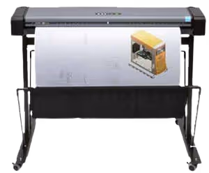
2D Scanners
Contex 2D Scanners
As a company, Contex delivers the most consistent, high-quality, fast 2D scanners on the market. That’s the reason why we refuse to sell any 2D scanners that aren’t Contex.
Wide Range of Cutting-Edge 3D Printers and Scanners
Elevate your design and manufacturing process with high-quality 3D printers and scanners
We offer a wide selection of state-of-the-art 3D printers and scanners to meet your design and manufacturing needs. Whether you're looking to bring your digital designs to life or capture real-world objects with precision, our range of products will empower you to innovate and create like never before.
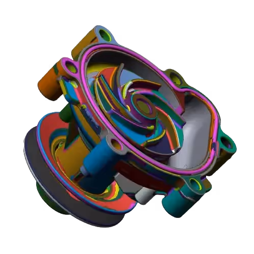
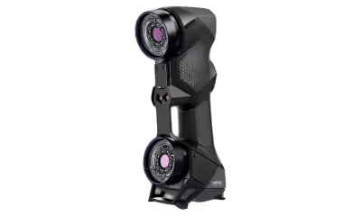
3D Scanners
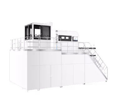
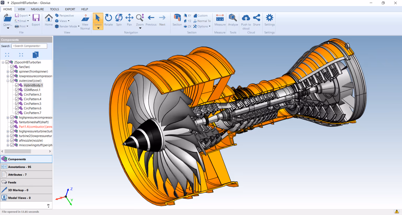
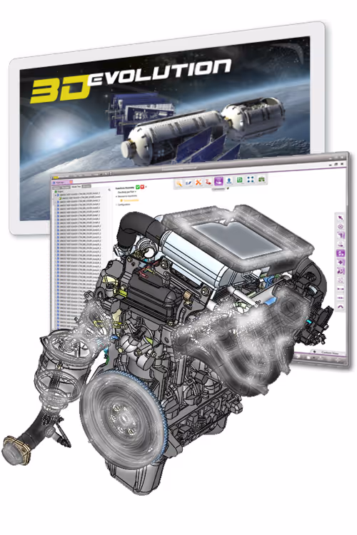
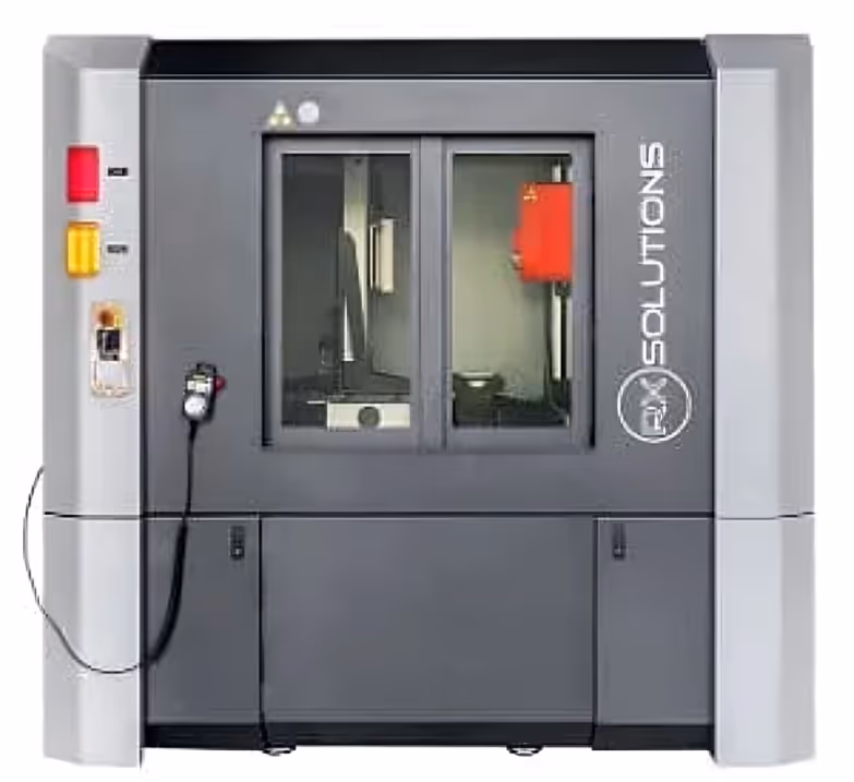
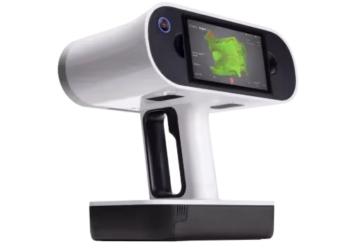
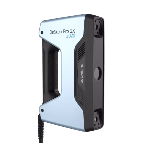
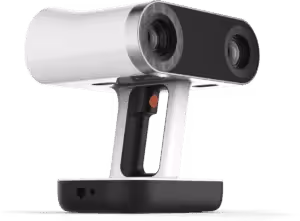
With over 30 years of scanning expertise, we bring you the finest selection of 3D scanners in the market. Our range includes top-of-the-line engineering scanners that deliver exceptional accuracy and efficiency.
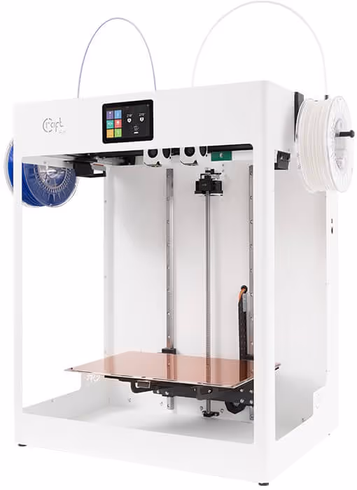
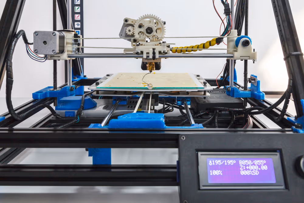
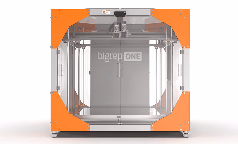
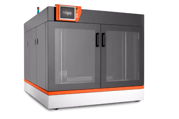
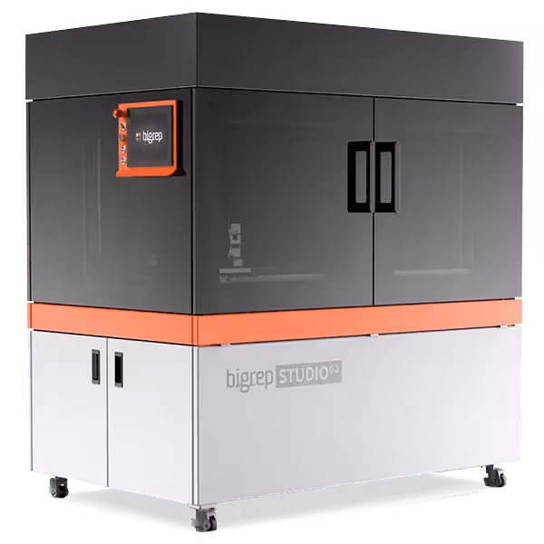
Discover our extensive collection of 3D printers that enable you to turn your digital designs into tangible objects. From small-scale prototypes to complex industrial parts, our printers provide the accuracy, speed, and versatility required for a wide range of applications. With cutting-edge features and user-friendly interfaces, our 3D printers make additive manufacturing accessible to businesses of all sizes. Check our entire selection of 3D printers here.
With over 30 years of scanning expertise, we bring you the finest selection of 3D scanners in the market. Our range includes top-of-the-line engineering scanners that deliver exceptional accuracy and efficiency.


-p-500.avif)